

Best Foods for Strong Bones & Joints: A Diet Guide
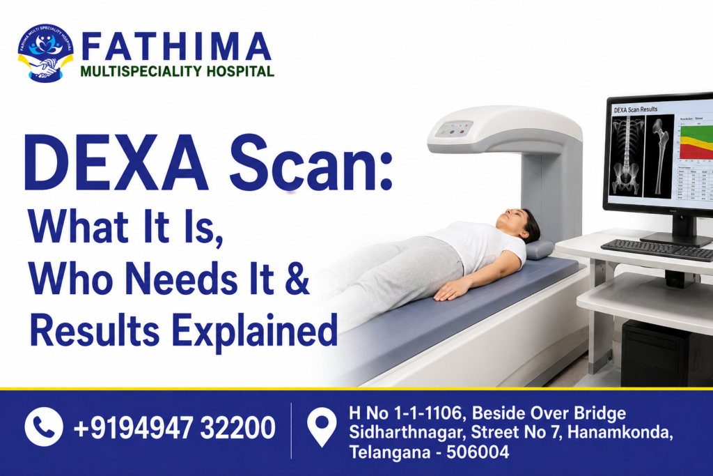
A DEXA Scan (Dual-Energy X-ray Absorptiometry) is a fast, painless, low-radiation imaging test that measures bone mineral density (BMD). It is the gold-standard diagnostic tool to detect osteoporosis, low bone mass (osteopenia), and predict fracture risk — typically completed in just 10–20 minutes.
Scan Duration: 10–20 min
Radiation Dose: Very Low
Injections / Prep: None
Primary Sites: Spine & Hip
DEXA stands for Dual-Energy X-ray Absorptiometry. It uses two low-dose X-ray beams at different energy levels to measure how dense your bones are. The denser your bones, the stronger and less fracture-prone they are. The DEXA scanner calculates the amount of X-ray energy absorbed by bone versus soft tissue, producing a precise Bone Mineral Density (BMD) score.
It is most commonly performed on the lumbar spine (lower back) and hip, as these are the sites most vulnerable to osteoporotic fractures. In some cases, the forearm or total body may also be scanned.
Unlike a traditional X-ray that only shows fractures after they occur, a DEXA Scan catches silent bone loss years before a fracture happens — making it a powerful preventive tool.
Osteoporosis is often called the “silent disease” because bone loss happens gradually — with no pain, no warning signs — until a fracture occurs. In India alone, over 50 million people are affected by osteoporosis, yet a vast majority remain undiagnosed.
A DEXA Scan is critical because it:
The following individuals are strongly recommended to undergo a DEXA Scan:
ullamcorper mattis, pulvinar dapibus leo.
The procedure is remarkably straightforward and entirely non-invasive:
i. Preparation: No fasting required. You may be asked to avoid calcium supplements 24 hours before the scan. Wear comfortable, metal-free clothing.
ii. Positioning: You lie flat on a padded table. The scanner arm passes slowly over your body — you remain completely still but do not enter a tunnel or enclosed space.
iii. Scanning: The machine emits two low-energy X-ray beams that measure bone density at key sites — typically the lumbar spine and hip. The dose is extremely small, roughly equivalent to a few hours of natural background radiation.
iv. Results: The scan produces a T-score and Z-score, which are interpreted by your orthopaedic specialist.
Your result is given as a T-score — a comparison of your bone density to that of a healthy young adult. Here is how to interpret it:
T-Score Range | Diagnosis | What It Means | Status |
-1.0 and above | Normal | Healthy bone density | Normal |
-1.0 to -2.5 | Osteopenia | Low bone mass; monitor and act early | Monitor |
-2.5 and below | Osteoporosis | High fracture risk; treatment needed | High Risk |
Your doctor will also review your Z-score, which compares your bone density to others of the same age and gender — particularly useful for younger patients and to identify secondary causes of bone loss.
At Fathima Multispeciality Hospital, a DEXA Scan is a vital part of pre-surgical assessment for patients undergoing knee or hip replacement surgery. Bone quality directly affects how well an implant integrates, the risk of periprosthetic fractures, and long-term surgical outcomes.
Dr. Sukesh Reddy P, who leads our orthopaedic department, uses DEXA results to personalise surgical planning — including implant selection, bone augmentation decisions, and post-operative rehabilitation protocols. This is especially important for our advanced Robotic Knee Replacement procedures, where bone quality data contributes to the precision planning algorithms.
The frequency depends on your initial result and clinical risk:
Frequently Asked Questions (FAQ’s)
1Q: What is a DEXA Scan?
Ans: A DEXA Scan is a painless, low-radiation test that measures bone mineral density to detect osteoporosis and fracture risk in 10–20 minutes.
2Q: What is a normal DEXA Scan T-score?
Ans: A T-score above -1.0 is normal. Between -1.0 and -2.5 is osteopenia. Below -2.5 indicates osteoporosis.
H No 1-1-1106, Beside Over Bridge Sidharthnagar, Street No 7, Hanamkonda, Telangana - 506004