

Best Foods for Strong Bones & Joints: A Diet Guide
The most common orthopaedic diagnostic tests are X-ray, MRI, CT scan, DEXA scan, Ultrasound, Blood tests, and Bone Scan. Each test serves a different purpose — X-rays detect fractures, MRI evaluates soft tissues, DEXA scan measures bone density, and blood tests identify inflammatory or metabolic conditions. Your orthopaedic doctor will choose the right combination based on your symptoms.
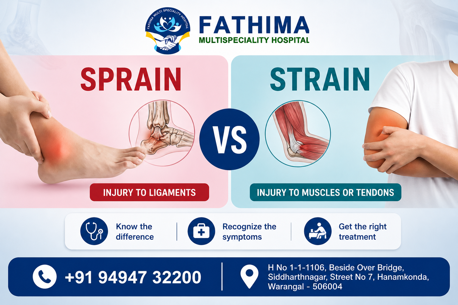
Bone and joint pain is one of the most common reasons people visit a doctor in India — and one of the most misdiagnosed. Pain in the knee could be arthritis, a ligament tear, a meniscus injury, or early osteoporosis. Pain in the back could be a disc problem, a fracture, or a nerve issue. Without the right diagnostic test, even the most experienced surgeon is working blind.
At Fathima Multispeciality Hospital, Warangal, Dr. Sukesh Reddy P (MS Orthopaedics) follows a structured, evidence-based diagnostic approach — ordering only the tests your specific condition truly needs. Here are the 7 essential orthopaedic tests and exactly what each one does.
i. X-Ray
Imaging · First-line test
Uses radiation to produce images of bone structure. Fast, affordable, and widely available.
FracturesArthritisDislocationsBone alignment
ii. MRI Scan
Imaging · Soft tissue specialist
Uses magnetic fields — no radiation. Gold standard for soft tissue evaluation.
Ligament tearsCartilage lossDisc herniationBone marrow
iii. CT Scan
Imaging · 3D bone detail
Combines X-ray with computer technology to produce cross-sectional 3D bone images.
Complex fracturesSurgical planningTumour detection
iv. DEXA Scan
Bone density · Osteoporosis test
Measures bone mineral density with minimal radiation. The gold-standard test for osteoporosis.
OsteoporosisFracture riskPre-surgery assessment
v. Ultrasound
Imaging · Soft tissue & joints
Uses sound waves — no radiation. Ideal for guiding injections and detecting fluid in joints.
Tendon tearsJoint effusionGuided injections
vi. Blood Tests
Lab · Inflammatory markers
Blood panels detect infection, inflammation, metabolic bone disease, and autoimmune arthritis.
ESR / CRPRheumatoid FactorUric AcidVitamin D
vii. Bone Scan
Nuclear imaging · Metabolic activity
A radioactive tracer is injected to identify areas of abnormal bone activity not visible on X-ray.
Stress fracturesBone infectionCancer spread
Your Symptom / Concern | Recommended Test | Why |
Suspected fracture after injury | X-Ray | Fast, accurate bone break detection |
Knee pain / suspected ligament tear | MRI Scan | Best for ACL, meniscus & cartilage evaluation |
Complex fracture, surgical planning | CT Scan | Detailed 3D bone geometry for surgeons |
Osteoporosis / fracture risk over 50 | DEXA Scan | Measures bone density with T-score |
Back pain / neck pain / nerve symptoms | MRI + X-Ray | Disc herniation, spinal cord, nerve compression |
Joint swelling, suspected arthritis | Blood Tests + X-Ray | Identifies inflammatory or metabolic cause |
Tendon or soft tissue injury | Ultrasound | Real-time soft tissue imaging without radiation |
Unexplained bone pain, suspected infection or tumour | Bone Scan + MRI | Detects abnormal metabolic bone activity |
At Fathima Multispeciality Hospital, Warangal, diagnostic tests are not just tick-box exercises — they are the foundation of every surgical and non-surgical treatment plan. Here is the structured pathway Dr. Sukesh Reddy P follows:
i. Clinical Examination First
Every patient undergoes a detailed physical examination before any test is ordered — joint range of motion, pain mapping, and neurological checks.
ii. First-Line Imaging (X-Ray)
An X-ray is taken in most cases to assess bone alignment, joint space, and rule out fractures or gross arthritis.
iii. Advanced Imaging if Required (MRI / CT)
Based on X-ray findings and clinical symptoms, MRI or CT is ordered to assess soft tissues or obtain 3D surgical planning data.
iv. DEXA Scan for Bone Density
All patients above 50, and those being evaluated for knee or hip replacement surgery, undergo a DEXA Scan for bone quality assessment.
v. Blood Tests for Inflammatory or Metabolic Conditions
ESR, CRP, Vitamin D, Uric Acid, and Rheumatoid Factor panels are ordered when arthritis, gout, or infection is suspected.
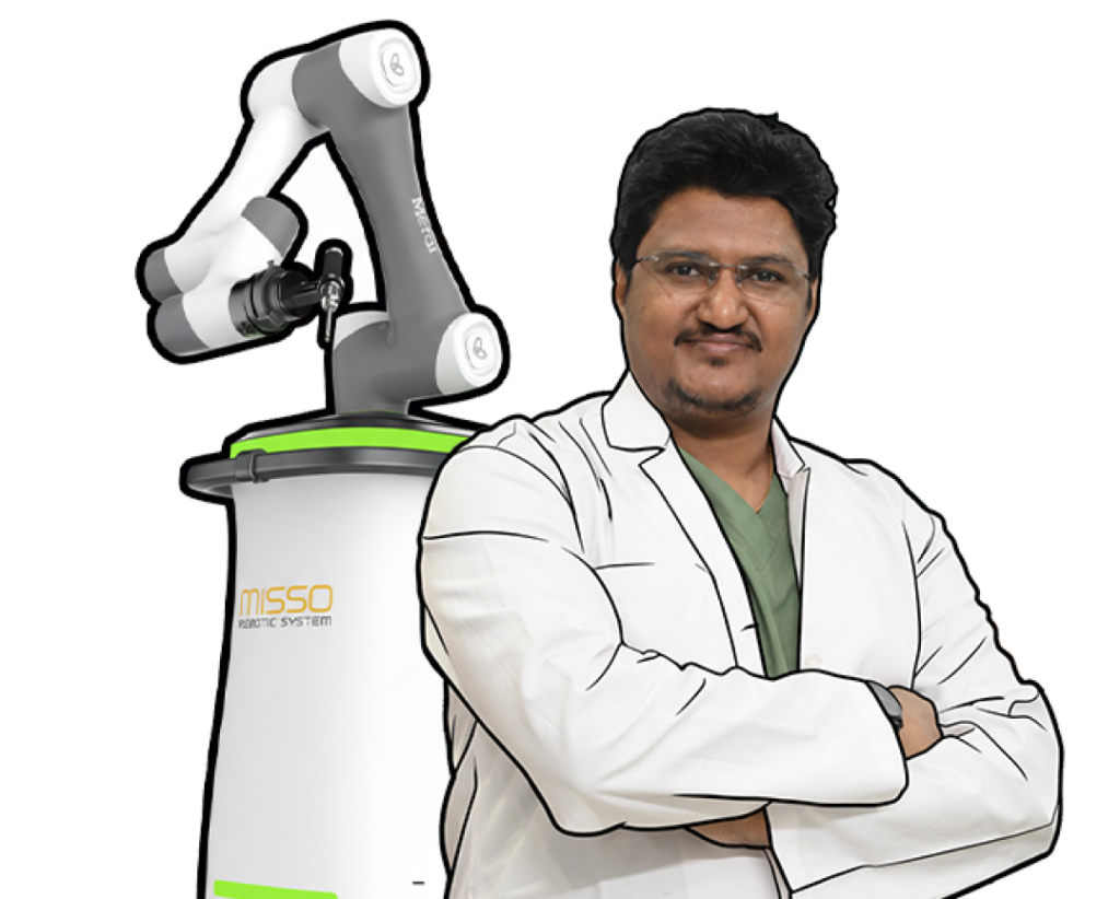
vi. Robotic Surgical Planning (Where Applicable)
For robotic knee or hip replacement, CT-based bone mapping feeds directly into the robotic system for precise, personalised implant positioning.
Do not self-diagnose. Never book diagnostic tests without consulting an orthopaedic specialist first. The wrong test wastes time and money — and can delay the correct treatment. Always get a clinical examination first.
H No 1-1-1106, Beside Over Bridge Sidharthnagar, Street No 7, Hanamkonda, Telangana - 506004